Projects
Community Resources
We are committed to fostering collaboration among labs studying the functional organisation of the plant cell nucleus and the 3D cytological organization of the genome. We promote knowledge transfer through publishing our tools and methodological developments and hosting inter-lab secondments for training.
These efforts were supported by the INDEPTH COST Action (CA16212, chair: Tatout C., co-chair: Baroux C.) that still bears legacy today. For more information, visit INDEPTH and see our final report, “INDEPTH: A Community Resource for Plant Sciences” as well as the compendium of Methods contributed by a large number of authors in the community “Plant Chromatin Dynamics: Methods and Protocols” (2018, doi: 10.1007/978-1-4939-7318-7) and its follow-up Edition (2024, ISBN 978-1-0716-4227-6, in press), and other references below.
Protocols
- Kracik-Dyer, E. and Baroux, C. , 3D STED Imaging of Isolated Arabidopsis thaliana Nuclei. Methods in Mol. Biol. 2024 in press
- Schubert, J., et al., A procedure for Dex-induced gene transactivation in Arabidopsis ovules. Plant Methods, 2022. 18(1): p. 41 DOI: 10.1186/s13007-022-00879-x.
- She, W., C. Baroux, and U. Grossniklaus, Cell-Type Specific Chromatin Analysis in Whole-Mount Plant Tissues by Immunostaining. Methods Mol Biol, 2018. 1675: p. 443-454 DOI: 10.1007/978-1-4939-7318-7_25.
- Fabrice, T.N., et al., Transmission Electron Microscopy Imaging to Analyze Chromatin Density Distribution at the Nanoscale Level. Methods Mol Biol, 2018. 1675: p. 633-651 DOI: 10.1007/978-1-4939-7318-7_34.
- Mendocilla-Sato, E., W. She, and C. Baroux, 3D Imaging of Whole-Mount Ovules at Cellular Resolution to Study Female Germline Development in Rice. Methods Mol Biol, 2017. 1669: p. 37-45 DOI: 10.1007/978-1-4939-7286-9_3.
- She, W., D. Grimanelli, and C. Baroux, An efficient method for quantitative, single-cell analysis of chromatin modification and nuclear architecture in whole-mount ovules in Arabidopsis. J Vis Exp, 2014(88): p. e51530 DOI: 10.3791/51530.
- Raissig, M.T., et al., Efficient and rapid isolation of early-stage embryos from Arabidopsis thaliana seeds. J Vis Exp, 2013(76) DOI: 10.3791/50371.
Tutorials for image analysis

- Randall, R.S., et al., Image analysis workflows to reveal the spatial organization of cell nuclei and chromosomes. Nucleus, 2022. 13(1): p. 277-299 DOI: 10.1080/19491034.2022.2144013.
- Mendocilla-Sato, E. and C. Baroux, Analysis of 3D Cellular Organization of Fixed Plant Tissues Using a User-guided Platform for Image Segmentation. Bio Protoc, 2017. 7(12): p. e2355 DOI: 10.21769/BioProtoc.2355.

Tools for image data analysis
We developed tools for interactively plotting and analysing segmented image data
- DataViz: reads data from segmented images containing spots and surfaces objects, different channels, and provide an interactive interface for customised plotting. See here: https://github.com/barouxlab/DataViz and doi:10.1080/19491034.2022.2144013
- ChromNano: analyses chromatin density distribution from TEM or dSTORM images. See here: https://github.com/barouxlab/ChromDensityNano and doi:10.1007/978-1-4939-7318-7_34
- OvuleViz: reads data from segmented images containing labelled cells objects, and provide an interactive interface for customized plotting. See here: https://github.com/barouxlab/OvuleViz and doi: 10.7554/eLife.66031
Community Reviews
- Dumur, T., et al., Probing the 3D architecture of the plant nucleus with microscopy approaches: challenges and solutions. Nucleus, 2019. 10(1): p. 181-212 DOI: 10.1080/19491034.2019.1644592.
- Baroux, C. and V. Schubert, Technical Review: Microscopy and Image Processing Tools to Analyze Plant Chromatin: Practical Considerations. Methods Mol Biol, 2018. 1675: p. 537-589 DOI: 10.1007/978-1-4939-7318-7_31.
- Tatout, C., et al., The INDEPTH (Impact of Nuclear Domains on Gene Expression and Plant Traits) Academy: a community resource for plant science. J Exp Bot, 2022. 73(7): p. 1926-1933 DOI: 10.1093/jxb/erac005.
Funding sources
Swiss National Science Foundation (SNSF), University of Zürich, Velux Foundation, Baumgarten Stiftung, Agence Nationale pour la Recherche (ANR)
Plant linker Histones
Linker histones, particularly H1, are essential components of chromatin that play a crucial role in the regulation of gene expression and genome organization. Structurally, linker histones consist of a central globular domain flanked by a long C-terminal tail and a shorter N-terminal extension, allowing them to bind to the nucleosome and the DNA linker regions between nucleosomes. This binding facilitates the compaction of chromatin into higher-order structures, influencing accessibility for transcription factors and other regulatory proteins. Linker histones are known to modulate chromatin dynamics, thereby impacting processes such as transcriptional repression and activation. In Arabidopsis, research has revealed a complex interplay of linker histones with various epigenetic marks, influencing developmental processes and stress responses.
|
|
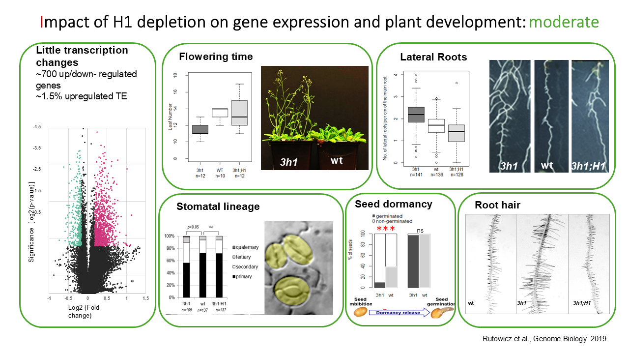
We showed that H1 linker histones are crucial for maintaining heterochromatic domains compaction, but dispensable for silencing at most TE loci. H1 deficiency also affects euchromatin structure and nucleosome distribution but this led to the deregulation of only a modest number of loci (~700), at least in controlled growth conditions. H1 depletion is not lethal for Arabidopsis and mildly affects some developmental processes such as seed dormancy, flowering time, and lateral root formation. We are currently investigating further the role of linker histones in physiological and developmental processes involving transcriptome reprogramming. |

| Additionally, we found that the transition from somatic to reproductive cell fate in female and male spore mother cells involves extensive chromatin reorganisation, characterized by heterochromatin decondensation and global changes in histone modifications (H3 methylation and acetylation) as well as reduced transcriptional activity. These events, which we found to be associated with gametophytic competence, are all preceded by linker histone depletion. We created point mutant variants of H1.1 and conditional mutants leading to the suppression of H1 eviction that now help us to elucidate the mechanisms of H1 eviction and roles in gametogenesis. |
|

- Rutowicz K, Lirski M, Mermaz B, Teano G, Schubert J, Mestiri I, Kroten MA, Fabrice TN, Fritz S, Grob S et al. 2019. Linker histones are fine-scale chromatin architects modulating developmental decisions in Arabidopsis. Genome Biol 20: 157.
- She W, Baroux C. 2015. Chromatin dynamics in pollen mother cells underpin a common scenario at the somatic-to-reproductive fate transition of both the male and female lineages in Arabidopsis. Frontiers in plant science 6: 294.
- She W, Grimanelli D, Rutowicz K, Whitehead MW, Puzio M, Kotlinski M, Jerzmanowski A, Baroux C. 2013. Chromatin reprogramming during the somatic-to-reproductive cell fate transition in plants. Development 140: 4008-4019.
- Teano G, Concia L, Wolff L, Carron L, Biocanin I, Adamusova K, Fojtova M, Bourge M, Kramdi A, Colot V et al. 2023. Histone H1 protects telomeric repeats from H3K27me3 invasion in Arabidopsis. Cell Rep 42: 112894.
Funding
Swiss National Science Foundation (SNSF), University of Zurich
Plant Chromatin Dynamics at Nanoscale

The chromatin organization in plant cells is highly dynamic, responding to both developmental cues and environmental factors. While much of our understanding comes from molecular profiling of the epigenome and 3D genome interactions, cytological studies also reveal significant changes in chromatin distribution. For example, heterochromatin, visible as chromocenters, fluctuates in quantity and location during developmental transitions such as germination, flowering, reproduction, and senescence, as well as in response to environmental factors like light, temperature, and pathogens. These shifts are often linked to changes in nuclear size and shape. However, we may be looking at only the surface. Less is known about the cytological distribution of euchromatin, which includes transcriptionally active regions, facultative heterochromatin, and various nuclear bodies. The 3D organization of euchromatin remains poorly understood. To explore the functional principles of chromatin organization at the nanoscale, our recent work has focused on optimizing workflows for sample preparation, immunolabeling/FISH, high-to-super-resolution 3D imaging, and quantification (see Community Resources). Key to our work are approaches of segmentation and spatial measurements enabling to quantify chromatin domains, their size, shape, position and abundance. We are now investigating the 3D nanoscale organization of epigenetic (H3K4me3, H3K27me) and transcription (RNA Pol II) domains in the nucleus during key cellular transitions, such as the dark-to-light transition in photomorphogenesis and the leaf-to-protoplast transition, marking dedifferentiation.
- Rutowicz K., Lüthi J., de Groot R., Holtackers R., Yakimovich Y., Pazmiño D. M., Gandrillon O., Pelkmans L., Baroux C. (2024) Multiscale chromatin dynamics and high entropy in plant iPSC ancestors. Journal of Cell Science, DOI: 10.1242/jcs.261703
- Kracik-Dyer, E., Baroux, C. (2025). 3D STED Imaging of Isolated Arabidopsis thaliana Nuclei. In: Baroux, C., Tatout, C. (eds) Methods for Plant Nucleus and Chromatin Studies. Methods in Molecular Biology, vol 2873. Humana, New York, NY.
- Randall, R. S., Jourdain, C., Nowicka, A., Kaduchová, K., Kubová, M., Ayoub, M. A., … Baroux, C. (2022). Image analysis workflows to reveal the spatial organization of cell nuclei and chromosomes. Nucleus, 13(1), 279–301.
- Baroux, C., Schubert, V. (2018). Microscopy and Image Processing Tools to Analyze Plant Chromatin: Practical Considerations. In: Bemer, M., Baroux, C. (eds) Plant Chromatin Dynamics. Methods in Molecular Biology, vol 1675. Humana Press, New York